Facility
Preclinical 7-tesla MRI system
One of the major features of MR is its non-invasiveness to living organisms at the time of measurement, which makes it possible to obtain information on various living organisms simply by lying down on the magnet. This feature makes it possible to apply the measurement methods developed and studied using the experimental equipment to clinical practice in a relatively short period of time. We have introduced the mini-7T device for animal experiments and are developing various methods to measure biological information in order to bridge the gap between animal experiments and clinical research.
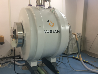
Fig. 1 Preclinical 7-tesla MRI scanner
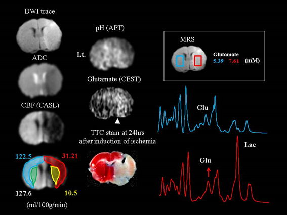
Fig. 2: Brain dynamics during middle cerebral artery occlusion in mice
DWI was measured at two B-values (B=1400 and 40 mm2/sec) by the spin echo method, while CBF was quantified by CASL. In addition, CEST, a semi-quantitative method to measure the exchange of protons between water molecules and amino, amide, and hydroxyl groups in brain tissue, is also available for imaging brain pH and glutamate maps.
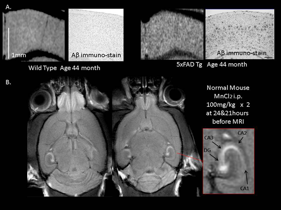
Fig. 3 MR microsopy
A. Senile plaque image of a 5XFAD Alzheimer Tg mouse (right) and an age-matched litter mate mouse (left), and Aβ immunostaining of the corresponding region. In the 5XFAD mouse, the aged mice is depicted as low-signal (black) by susceptibility. Manganese permeates calcium channels and is taken up into neurons according to neuronal activity, shortening the T1 time. DG: dentate gyrus.
1-757 Asahimachi-dohri, Niigata, Niigata, Japan 951-8585
Copyright c Center for Integrated Human Brain Science, University of Niigata.
