Facility
MR Scanners for human
(1)Horizontal 7T human MRI system (7T)
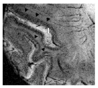
One of the final goals for clinical imaging research is the development of a non-invasive system capable providing images of microscopic resolution. With 3T systems, we have achieved routine clinical imaging with gross pathological resolution. However, it became apparent that yet higher S/N would be necessary to develop realistic human magnetic resonance microscopy. Subsequently, a system with a 7T magnet was installed in April 2003. MR microscopic SWI image of a patient with Alzheimer’s disease is presented below.
(2)Horizontal 3T human MRI system (H3T)
This horizontal system is the first 3T system installed in Japan in April 1996 suitable for human imaging. It is capable of performing a variety of non invasive magnetic functional imaging. These includes functional MR imaging, diffusion tensor analysis as well as Magnetic Resonance Spectroscopic Imaging. A number of intra and extra mural collaborated projects are on going.
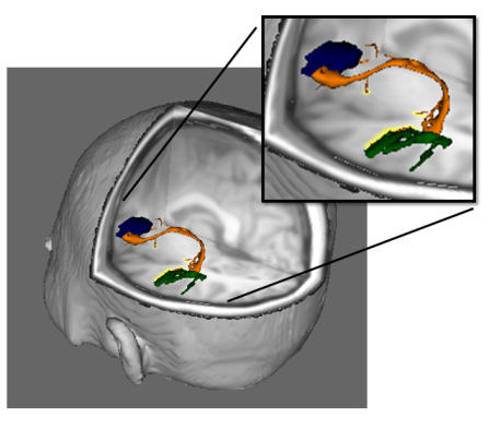
An example of probabilistic tractography, a tract (orange) connecting motor (blue) and sensory (green) language areas identified by fMRI. Probabilistic tractography is delineating neuronal tracts by Bayesian statistics using eigenvector information obtained by diffusion tensor analysis of MRI diffusion-weighted images.
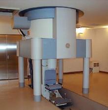
(3)Vertical 3T human MRI system (V3T)
The first human vertical system with a 3T large bore vertical magnet, was constructed in January 1999. in our system, the subject sits upright on chair, which is elevated into the magnet by electronic actuators. The subject may drive a car, perform the musical instruments such as the piano or undergo variety of other experiences not possible with current horizontal bore systems. This vertical system provides information regarding gravitational effects on regional perfusion and evaluation of gravitational effects in those patients with brain ischemia, et al.
1-757 Asahimachi-dohri, Niigata, Niigata, Japan 951-8585
Copyright c Center for Integrated Human Brain Science, University of Niigata.
